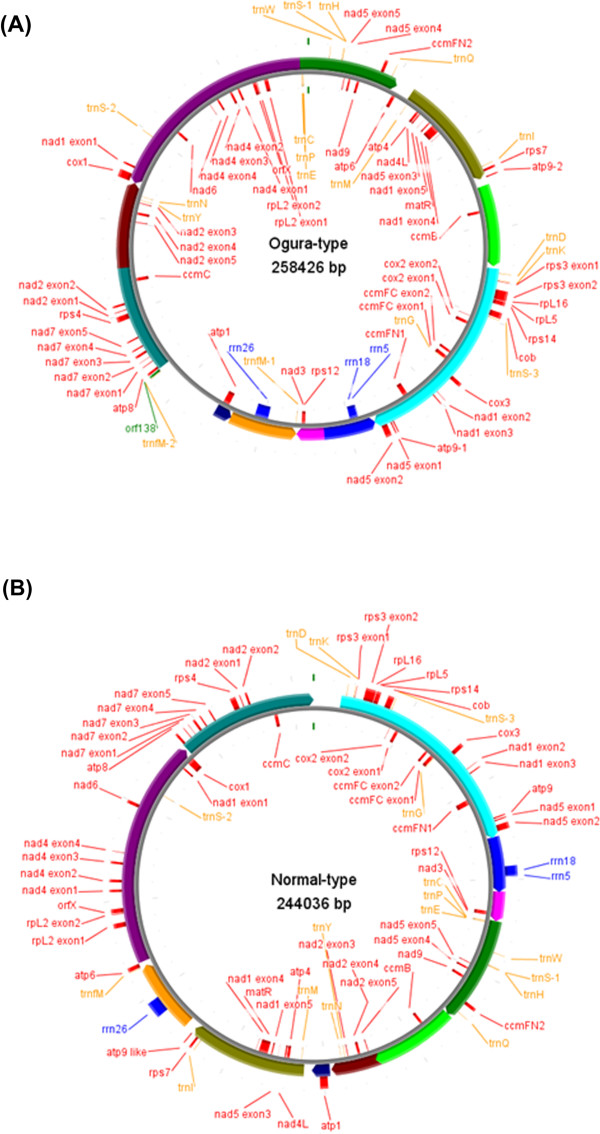
Figure 1.
The organization of Ogura-type (A) and normal-type (B) mitochondrial genomes represented as a "master circle". Features on forward and reverse strands are drawn on the outside and inside of the circles, respectively. Protein-coding genes are shown in red, rRNAs in blue, tRNAs in orange and orf138 in lime green. The arcs in the same colors indicate syntenic regions between Ogura- and normal-type genomes (refer to Figure 2). Genome maps were made with CGviewer [49].

