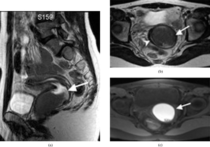
Figure 6.
A 19-year-old woman with didelphys uterus (Class III). (a) Sagittal T2 weighted MR image of a didelphys uterus with a transverse vaginal septum causing unilateral haematocolpos (arrow). (b) Axial T2 weighted MR image shows a dilated left vaginal canal with predominantly low signal intensity fluid (arrow) and compressed right vaginal canal (arrowhead). (c) Correlating axial T1 weighted fat saturation MR image shows hyperintense fluid in the distended left vaginal canal in keeping with haematocolpos (arrow).

