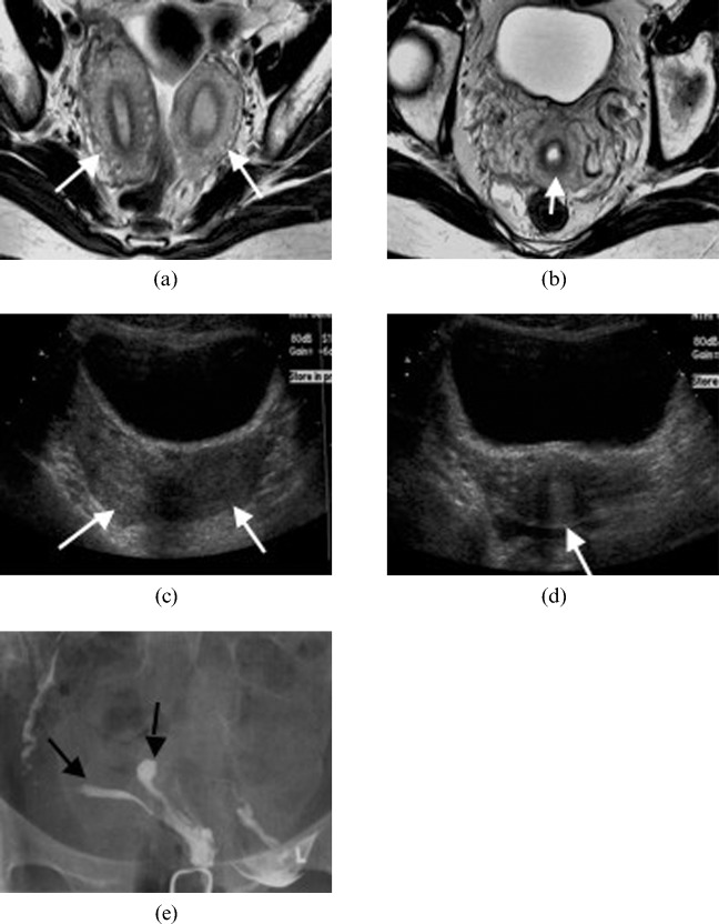
Figure 7.
A 30-year-old woman with a bicornuate uterus (Class IV). Axial T2 weighted image illustrating (a) two separate uterine horns (arrows) that fuse at its inferior end to give (b) a single cervix (arrow). Transverse ultrasound images demonstrating (c) two uterine horns (arrows) and (d) a single cervix (arrow). (e) Hysterosalpingography of a bicornuate uterus. The contrast material fills two separate uterine horns (arrows).

