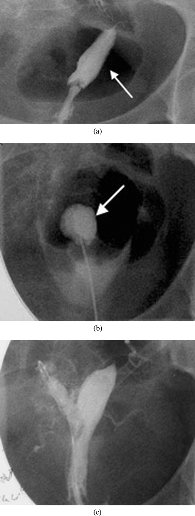
Figure 14.

A 22-year-old with intense menstrual pain who was investigated and found to have a bicornuate uterus with a non-communicating right horn. (a) Hysterosalpingogram reveals filling of the left horn (arrow) but not the obstructed right horn. (b) Intraoperative hystosalpingogram illustrating the puncture through the uterine septum into the obstructed right horn (arrow). This was performed following needle puncture of the septum with ultrasound guidance. A wire was then placed into the cavity and the gynaecologist then used the wire to guide resection of part of the wall of the obstructing right horn. (c) Subsequent hysterosalpingogram shows filling of the previously obstructed right horn. Note that spasm of the right tube prevented uniform tubal filling. Subsequent selective salpingography demonstrated the right tube to be patent. The patient had resolution of her pain and was very pleased at 2 year follow-up.
