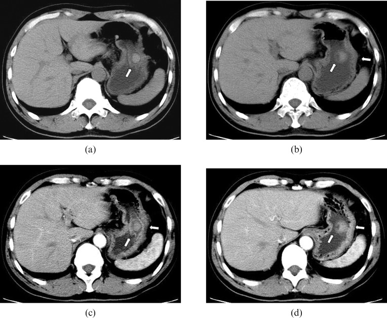
Figure 2.
CT findings. (a, b) Pre-enhanced CT scan shows a round mass of slight higher density with a depression on its cupula (arrows); the mass is 1.4 × 1.5 cm in size and has a CT value of 51.1–56.3 HU. (c, d) 60 s after intravenous contrast injection, the lesion shows slight homogeneous enhancement with an increase in CT value to 62.1–69.8 HU, and the depression in the lesion is clearer. The serous membrane of the stomach has not been involved (arrows).

