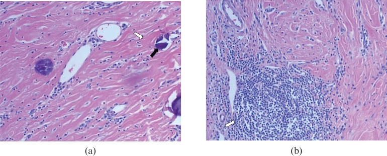
Figure 3.
Histological findings (haematoxylin and eosin staining (×200). (a) Hyalinised collagenous tissue (white arrow) with psammomatous calcifications (black arrow) is clearly revealed. (b) Dense hyalinised collagenous tissue and focal lymphocytic infiltration with lymphoid follicle formation (arrow) are demonstrated.

