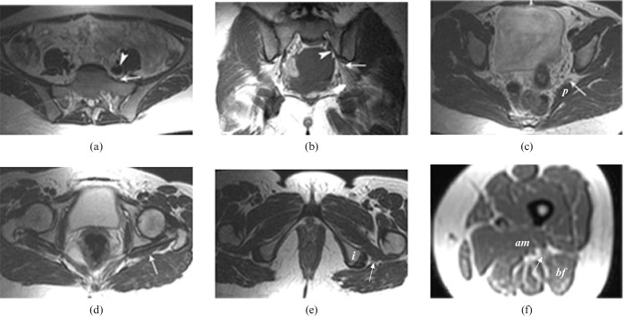
Figure 1.
Normal MRI appearance of the lumbosacral trunk, sacral plexus and sciatic nerve. Axial T1 weighted image (a) at the level of the sacral wings shows the lumbosacral trunk (arrow) anterior to the sacrum and posterior to the iliac vessels (arrowhead). Coronal T1 weighted image (b) shows the normal sacral plexus (arrowhead) passing anterior to the sacral wings and continuing inferolaterally to leave the pelvis through the greater sciatic foramen (thin arrow) as the sciatic nerve (thick arrow). Axial T2 weighted image (c) at the level of the greater sciatic foramen shows the sacral plexus (arrow) anterior to the piriformis (P) muscle and lateral to the inferior gluteal vessels. Axial T2 weighted image (d) at the level of the gluteal region shows the sciatic nerve (arrow) superficial to the lateral rotator muscles and deep to the gluteus maximus muscle. Axial T2 weighted image (e) at the level of the infragluteal region shows the sciatic nerve (arrow) between the ischial tuberosity (i) and the greater trochanter. Axial T1 weighted image (f) at the mid-femoral level shows the sciatic nerve (arrow) between the adductor magnus (am) and biceps femoris (bf) muscles.

