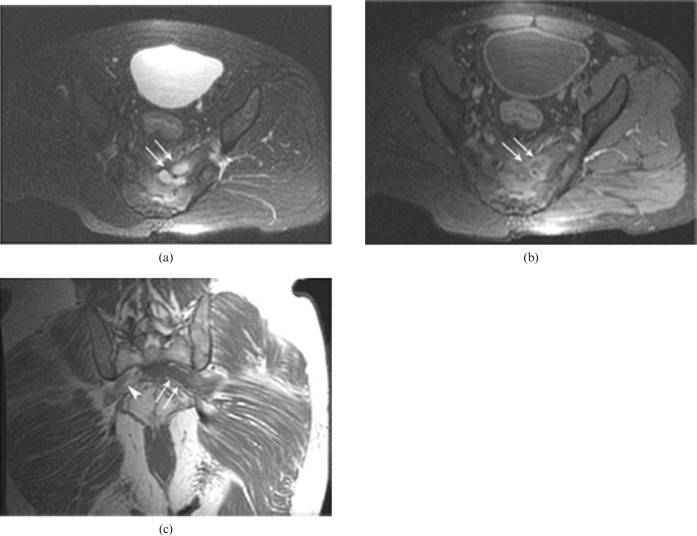
Figure 7.
Presacral abscess in a 55-year-old male patient with a left lumbosacral plexopath. A contrast-enhancing signal increase consistent with sacral osteomyelitis is observed on the fat suppression axial T1 weighted images following intravenous Gd administration (a) and on the axial fat suppression T2 weighted images (b). In addition, a presacral region cystic mass affecting the left lumbosacral plexus and consistent with an abscess is seen, demonstrating peripheral contrast attenuation (arrows). The right lumbosacral plexus appears normal (arrowhead) on the coronal T1 weighted images (c). The presacral abscess effect on the left lumbosacral plexus is clearly observable (arrows).

