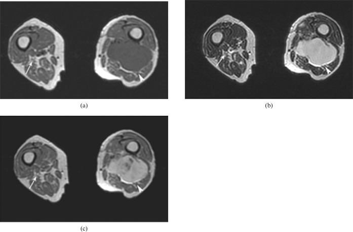
Figure 12.
Soft-tissue sarcoma in a 39-year-old female patient with left-side sciatica. Axial T1 weighted (a), T2 weighted (b) and T1 weighted images following intravenous contrast administration (c) demonstrating a soft-tissue mass with significant contrast enhancement in the posterior femoral compartment; the close relation of the mass to the sciatic nerve is seen (arrowheads). The right sciatic nerve appears normal (arrows).

