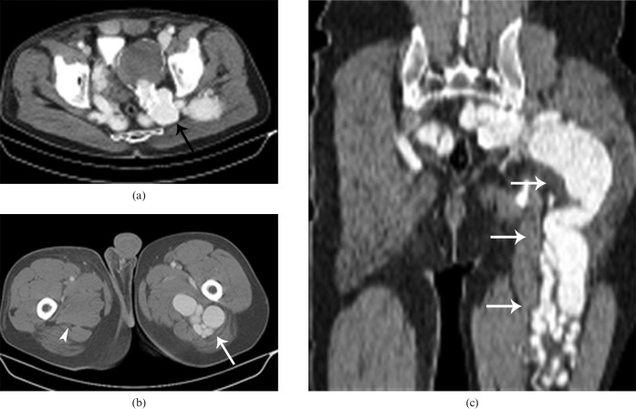
Figure 19.
Arteriovenous fistula in a 49-year-old male patient with left sciatica and a history of penetrating trauma 2 years previously. Axial (a,b) and coronal reformatted (c) CT images demonstrating a thrombosed aneurysm (black arrow) and diffuse varicose veins (white arrows) related to the arteriovenous fistula pressing on the left lumbosacral plexus and sciatic nerve. The right sciatic nerve appears normal (arrowhead).

