Figure 1.
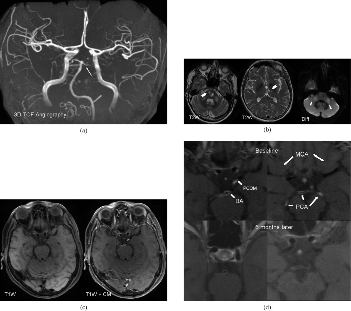
(a) Three-dimensional time-of-flight (TOF) MR angiography. The arrow points to lumen irregularities in the basilar artery and the posterior cerebral artery. These findings are unspecific and can be found in atherosclerotic disease as well as in central nervous system (CNS) arteritis. (b) Axial T2 weighted (T2W) MR images of the brain show hyperintense areas (chevron) in the left and right pons and in the left thalamus, consistent with ischaemic infarctions. The arrowheads point to hyperintense areas in both cerebellar peduncles, as an unspecific sign for cerebellitis. (c) Axial T1 weighted (T1W) images pre- and post-contrast show strong contrast enhancement in the basilar artery, both posterior cerebral arteries and in the left posterior communication artery, suggestive of CNS vasculitis. No contrast enhancement is seen in both middle cerebral arteries. CM, contrast media. (d) Magnified axial T1 weighted post-contrast images at baseline (first row) and 6 months after initiation of therapy (second row) show an absence of contrast enhancement in the left posterior cerebral artery (PCA) and in the left posterior communication artery (PCOM) and a decrease in the contrast enhancement in the basilar artery (BA) and the right PCA.

