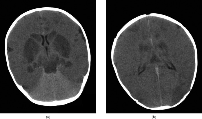
Figure 3.
Diffuse hypoxic brain injury. 3-month-old infant scanned post mortem after presenting following a history of overlying. Unenhanced cranial CT at (a) the basal ganglia and (b) the bodies of the lateral ventricles showing a global reduction in cerebral density with loss of cortical visualisation and a “bright cerebellum” sign, with more pronounced hypodensity of the deep grey matter nuclei, the hippocampi, the midbrain and parts of the cortex.

