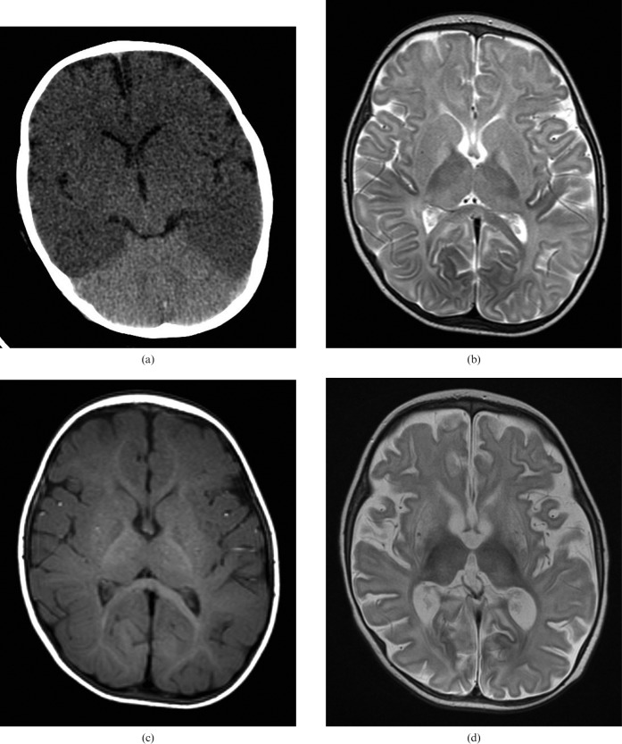
Figure 4.
Diffuse hypoxic brain injury in a 7-month-old infant admitted following a successful resuscitation of cardiac arrest with associated dilated cardiomyopathy. (a) Unenhanced cranial CT scan on the day of admission shows global reduction in cerebral density with reduced grey/white matter differentiation and a “bright cerebellum sign”. (b) Unenhanced axial T2 and (c) T1 weighted MRI scans 4 days following initial CT showing T2 hyperintensity in the globi pallidi, with subtle T2 hyperintensity in the putamen and thalami bilaterally. Note the absence of subdural haematoma on the T1 weighted image. (d) Axial T2 weighted MRI at 3 weeks following the acute presentation, demonstrating generalised atrophy, particularly affecting the basal ganglia.

