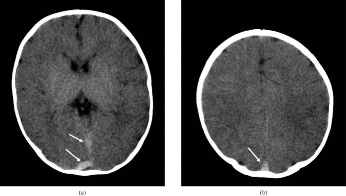
Figure 5.
Post-mortem cranial CT in a 3-month-old infant who presented to the emergency department in cardiorespiratory arrest after being found unresponsive in the parents' bed. Images (a) at the level of the basal ganglia and (b) through the centrum semiovale show generalised reduction in grey/white matter differentiation. Note the dense post-mortem appearances of the dural venous sinuses (white arrows).

