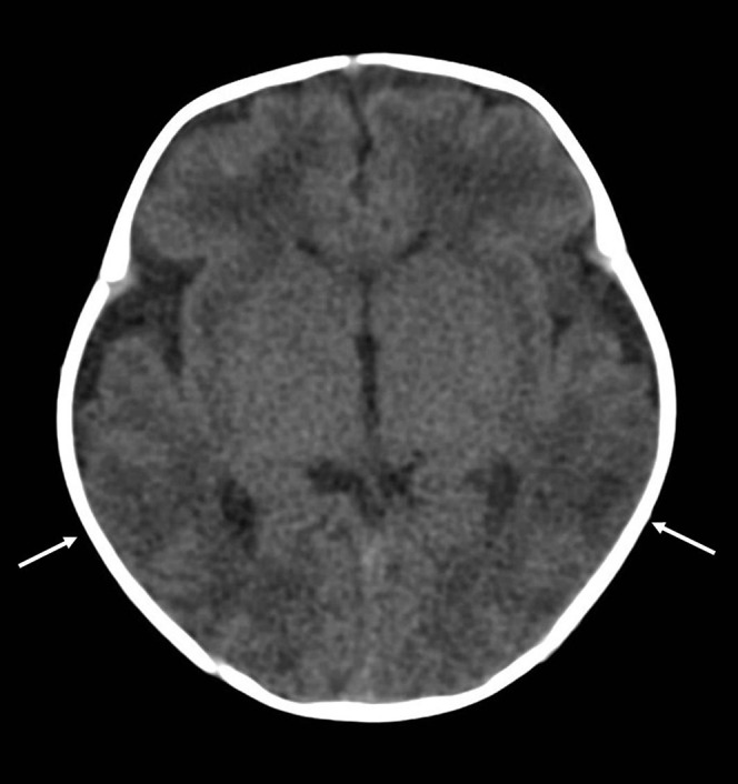
Figure 6.

Patchy hypoxic brain injury in a 1-month-old infant following an admission for apnoea. A cardiorespiratory arrest was witnessed on the ward. Unenhanced cranial CT scan shows focal loss of grey/white matter differentiation in the posterior temporal regions bilaterally (white arrows).
