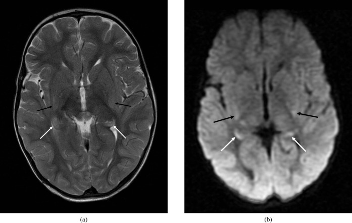
Figure 7.
Focal hypoxic injury in a child aged 1 year and 11 months. MRI scan performed 3 days after a near-drowning incident. (a) Axial T2 image shows subtle hyperintensity of the hippocampal tails (white arrows) and posterior putamen bilaterally (black arrows), with (b) corresponding high signal on the B = 1000 diffusion-weighted images.

