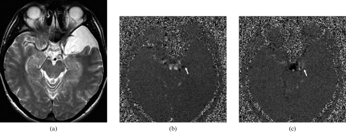
Figure 2.
A 42-year-old male with communicating left middle cranial fossa arachnoid cyst. (a) T2 weighted axial image shows left middle cranial fossa arachnoid cyst that is isointense to cerebrospinal fluid (CSF). CSF flow MRI (b) and (c) depict hypo- and hyperintensity (arrow), respectively, originating from suprachiasmatic cistern demonstrating communication between cyst and cistern. While, the pulsatile flow is seen only at the communicating site, it is not present throughout the cyst. This flow pattern is consistent with a communicating arachnoid cyst.

