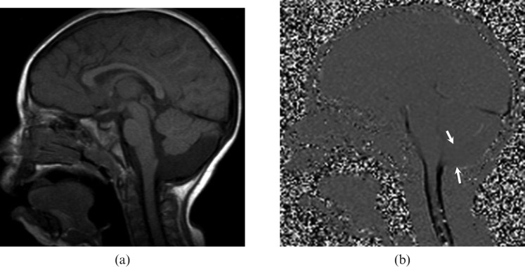
Figure 4.
A 2-year-old girl with a non-communicating posterior fossa arachnoid cyst. (a) T1 weighted sagittal image shows a retrocerebellar cystic lesion and scalloping in the occipital bone. (b) Cerebrospinal fluid (CSF) flow MRI reveals no communication between the cyst and the superior–posterior cervical subarachnoid space (arrows).

