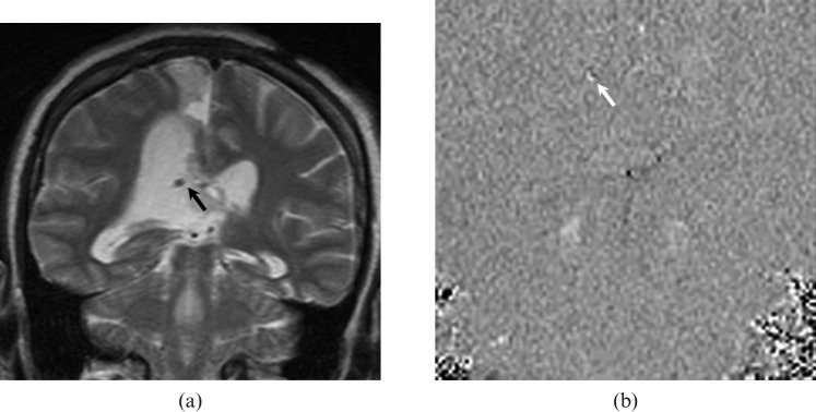
Figure 11.
A 34-year-old female patient with a ventriculoperitoneal shunt inserted for obstructive hydrocephalus. (a) Coronal T2 weighted MRI shows the ventriculoperitoneal shunt (black arrow) within the right ventricle. (b) Coronal cerebrospinal fluid flow MRI demonstrates brighter signal intensity (white arrow) than the background, suggesting patency at the site of the shunt. This flow pattern is consistent with the one-way flow in the ventriculoperitoneal shunt.

