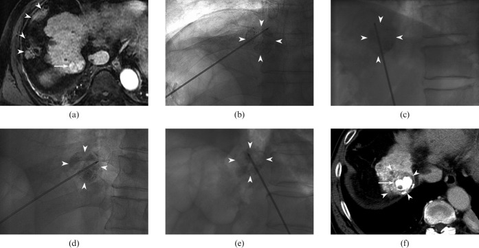
Figure 1.
64-year-old man with hepatocellular carcinoma (HCC) directly contacting the right hemidiaphragm (segment VIII). (a) Transverse arterial phase T1 weighted image (repetition time/echo time, 4.3/2.0 ms; flip angle, 12°) reveals a hyperintense lesion (arrow). Note that overlying colon (arrowheads) in the right perihepatic space precludes transverse approach of radiofrequency (RF) electrode to the index tumour. The tumour was invisible on ultrasound because of poor acoustic window. (b, c). 3 days after transarterial chemoembolisation (TACE), he underwent RF ablation under fluoroscopic guidance. Anteroposterior (b) and lateral (c) fluoroscopic views show obliquely angled approach of RF electrode into the index tumour with lipiodol retention (arrowheads) without traversing the thorax. (d, e) Overlapping treatments were performed in order to achieve adequate ablative margin. For the second ablation cycle, electrode placement was adjusted to cover a different portion (d and e) of the index tumour (arrowheads). (f) Transverse portal phase CT image obtained immediately after ablation shows index tumour (arrow) surrounded by non-enhancing ablative zone (arrowheads).

