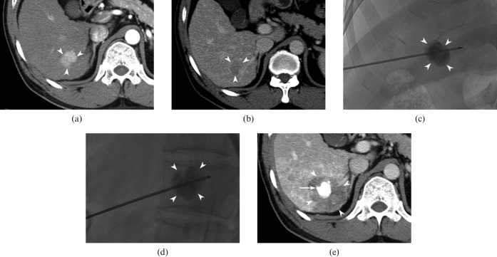
Figure 2.
46-year-old man with hepatocellular carcinoma (HCC) in the liver. (a, b) Transverse CT scan shows a 2.3 cm nodule (arrowheads) in segment VI that is hypervascular in the arterial phase (a) and washes out in the delayed phase (b), consistent with HCC. (c, d) 2 days after transarterial chemoembolisation (TACE), he underwent radiofrequency (RF) ablation. Anteroposterior (c) and lateral (d) fluoroscopic images show the RF electrode properly placed through the centre of the index tumour (arrowheads). (e) Transverse portal phase CT image obtained immediately after ablation reveals that the index tumour with retained iodised oil (arrow) is ablated with a sufficient ablative margin (arrowheads).

