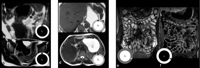
Figure 2.
Different oral contrast agents in MRI of the small intestine (SI). Double-negative contrast agents produce low-signal intensity of bowel lumen on both (a) T1 and (b) T2 weighted sequences. With these contrast agents the visualisation of the bowel's wall (w) is preferred to that of the lumen (l), emphasising the identification of parietal and extra-parietal findings such as oedema or fat stranding. With the use of positive contrast agents, the visualisation of bowel lumen is privileged on both (c) T1 and (d) T2 weighted sequences; the most significant limitation is represented by the fact that wall enhancement after intravenous Gd administration can be masked by the higher lumen signal on T1 weighted sequences. Biphasic contrast agents produce an optimal contrast between lumen and walls on both (e) T2 weighted (bright lumen and dark walls) and (f) T1 weighted sequences (dark lumen and bright walls).

