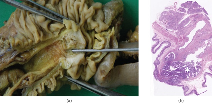
Figure 3.
Adenocarcinoma. (a) Photograph of resected and opened duodenum from a 67-year-old man who presented with melaena shows a 2.0 cm vegetating lesion originating from the mucosa surrounding the duodenal papilla. (b) Low-power photomicrograph (magnification ×4; haematoxylin–eosin stain) of the lesion shows neoplastic infiltration (asterisk) of the mucosal epithelium and muscularis propria.

