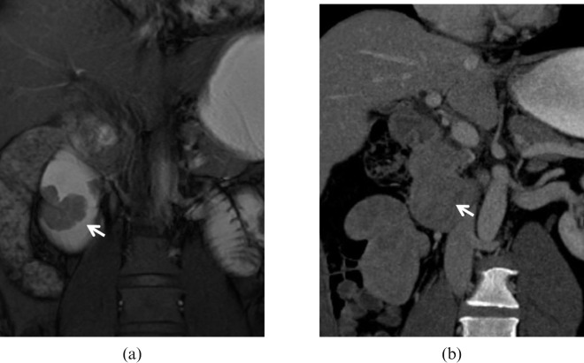
Figure 5.
Adenocarcinoma. 49-year-old male with melaena and endoscopic diagnosis of duodenal mass. The high intraluminal signal achieved on (a) MR true fast imaging with steady-state precession (true-FISP) image after water distension of the duodenum allows the identification of a polypoid lesion (arrow) with a thick stalk and lobulated profiles facing the duodenal papilla. The contour of the outer wall layers is preserved, excluding deep extraluminal infiltration. (b) In this case, the poor attenuation gradient and lesser lumen distension attained on multidetector CT prevent a precise delineation of the lesion (arrow).

