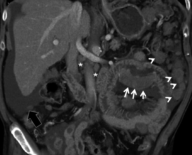
Figure 6.

Adenocarcinoma. 89-year-old male with weight loss, abdominal pain and melaena admitted to the emergency room. Multidetector CT images obtained after oral administration of a low-concentration contrast agent demonstrate an eccentric and partially stenosing wall thickening involving one of the proximal jejunal loops (arrows). The lesion extends beyond the wall infiltrating the perivisceral fat. Multiple peritoneal implants (arrowheads), perihepatic ascites (black arrow) and some centimetric lymphadenopaties (asterisk) are also visualised.
