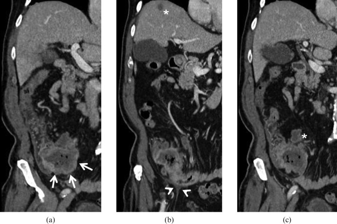
Figure 7.
Adenocarcinoma. 66-year-old male with weight loss, melaena and incomplete colonoscopy. Multidetector CT images demonstrate a large mass originating from the last ileal loop ((a) arrows) infiltrating the ileocaecal valve. The lesion extends into the perivisceral fat and infiltrates the peritoneum ((b) arrowheads). A small liver metastasis ((b) asterisk) and necrotic lymphadenopaties ((c) asterisk) can also be identified.

