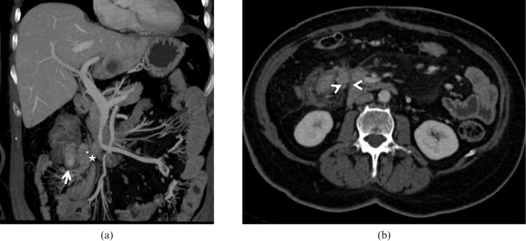
Figure 10.
Carcinoid tumour. 50-year-old male with multiple liver lesions identified at a routine ultrasound scan from an unknown primary cancer. (a) Multidetector CT images obtained in the portal-venous phase demonstrate an enhancing ileal lesion (arrow) with mass forming desmoplastic reaction in the mesentery (asterisk) the prominent mesenteric involvement is well visualised on the axial images ((b) arrowheads).

