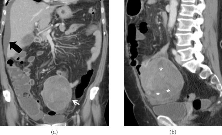
Figure 16.
Low-grade subserosal gastrointestinal stromal tumour (GIST). Same patient as Figure 15. (a) Multidetector CT images demonstrate a large lesion (arrow) with well-defined margins originating from the mesenteric side of one of the last ileal loops, perihepatic ascites is confirmed (black arrow). (b) Sagittal reconstruction clearly demonstrates the prevalent extraluminal extension of the lesion, also evidencing multiple necrotic foci inside the lesion (asterisk).

