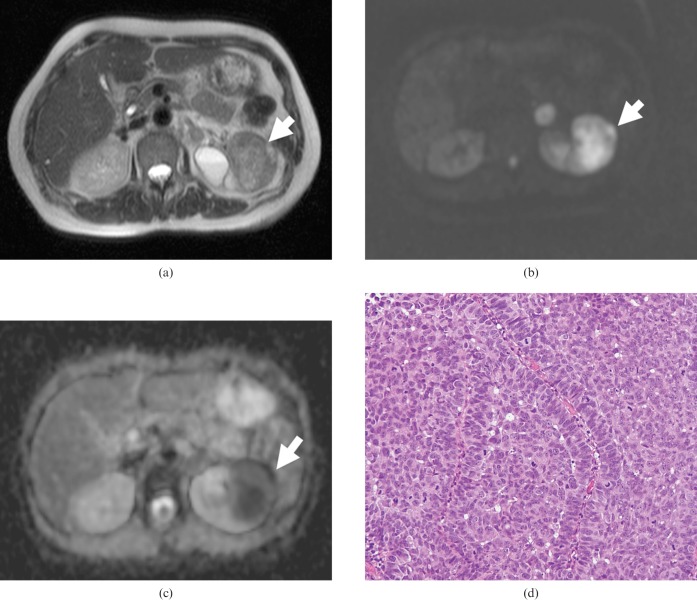
Figure 1.
Transverse MR images of a 65-year-old woman with transitional cell carcinoma of the left kidney. (a) T2 weighted image showing a low-signal-intensity mass of the left kidney (arrow). (b) On DWI, the tumour showed very high signal intensity (arrow). (c) The apparent diffusion coefficient map shows the low ADC value (0.71 × 10⁻³ mm2 s) of the tumour (arrow). (d) Haematoxylin and eosin (H&E) staining confirmed the histological diagnosis of TCC. Densely packed solid tumour cells with hypercellularity were seen (original magnification, ×100).

