Figure 1.
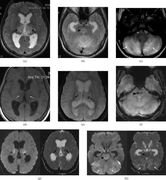
48-year-old male found comatose on admission with Glasgow coma scale (GCS) 4/15. Axial T2 weighted images showing multiple foci of hyperintensities in (a) bilateral thalami (black arrows); (b) brain stem (arrowhead); and (c) cerebellar hemispheres (white arrows). (d) Axial T1 weighted image showing corresponding hypointensities in the bilateral thalami (white arrows). Axial gradient-echo images show blooming artefacts in (e) bilateral thalami (black arrows) and (f) brain stem (arrowhead) compatible with petechial haemorrhages. Axial diffusion weighted images showing restricted diffusion in (g) bilateral thalami (black arrows), (h) brain stem (arrowheads) and cerebellar hemispheres (white arrows).

