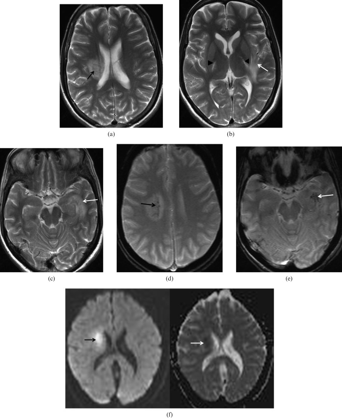
Figure 2.
A 32-year-old female presented with a history of fever, chills and rigors for the past 10 days. Axial T2 weighted images show multiple foci of hyperintensities in the (a) right corona radiata (black arrow), (b) posterior limb of bilateral internal capsule/thalami (arrowheads) and (c) left temporal lobe/hippocampus extending into the left insular cortex (white arrows). Axial gradient-echo images showing old haemorrhages in (d) right corona radiata (black arrow) and (e) left temporal lobe/hippocampus (white arrow). Axial diffusion-weighted images showing restricted diffusion in right corona radiata (arrows).

