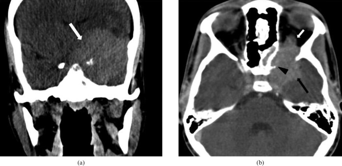
Figure 1.
(a) Non-contrast coronal CT scan shows a homogeneously hyperdense extra-axial lesion (white arrow). (b) Contrast-enhanced axial CT scan shows a homogeneously enhancing extra-axial lesion (black arrow), with extension into the orbit (white arrow) and erosion of left sphenoid wing (black arrow head).

