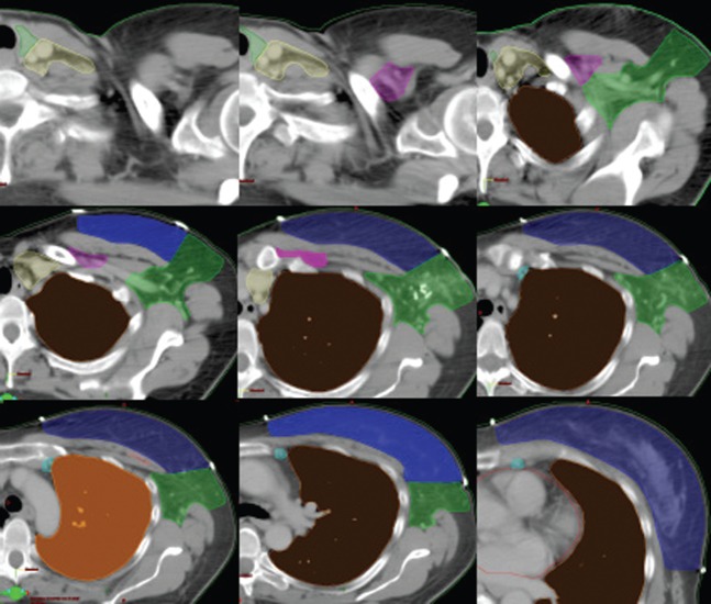
Figure 1.
Examples of delineation shown in the guide and simplified rules of delineation.
Supraclavicular region: contouring of the supraclavicular region is guided by the origin of the internal mammary artery.
Cranial: Thyroid cartilage
Caudal: Clavicular head
Medial (med): Trachea
Posterior (post)-lateral (lat): Anterior scalene muscle
Post-med: Carotid artery
Infra clavicular region: The infraclavicular region corresponds to the lymphatic drainage between axillary vertex, and the superior limit of the axillary LN dissection (LND).
Cranial: Pectoralis minor
Caudal: Sternoclavicular joint
Lat: Pectoralis minor (medial side)
Med: Clavicle
Ant: Pectoralis major
Post: Axillary artery
Internal mammary chain: The lymph nodes of the IMC are located within the anterior interspaces; they are located either medial or lateral to the vessels and are concentrated in the upper three interspaces.
Ant: Ant. part of the vascular area
Post: The pleura
Med: Medial limit of the vascular area
Lat: Lateral limit of the vascular area
Caudal: Superior side of the fourth rib
Cranial: Inferior limit of supraclavicular area
Rotter LN or intra pectoral node: situated between: pectoralis major and pectoralis minor at the second intercostal space
Axilla:
Ant: Pectoralis major and pectoralis minor
Post: Subscapularis, teres major and latissimus dorsi
Med: Seratus anteriorLat: 5 mm backward the skin
Caudal: fourth and fifth ribs
Cranial: Inferior limit of infraclavicular volume or “first clip”

