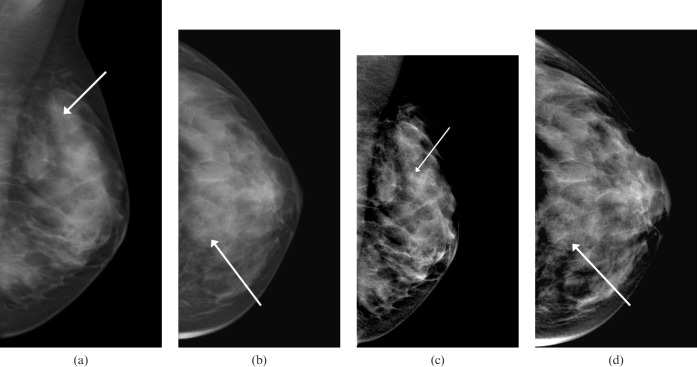
Figure 1.
A left-sided mammogram of a 45-year-old woman shows faint microcalcifications in the upper breast (arrows). (a,b) Images without Premium View show calcifications, but these are very faint and difficult to quantify. (c,d) The same mammographic images with Premium View applied show the calcifications to be much more contrasted, and easier to quantify in terms of appearance and extent. An increased soft tissue contrast in overlying breast tissue may allow the visibility of these to be further enhanced. These were subjected to a stereotactic biopsy, and high-grade ductal carcinoma in situ was obtained pathologically.

