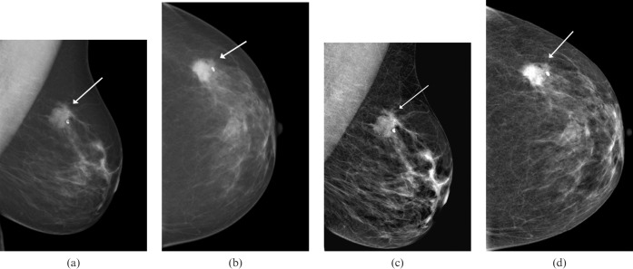
Figure 2.
A left-sided mammogram in a 65-year-old woman presenting with a left breast lump. (a,b) Images without Premium View show a highly suspicious lesion in the outer breast that is consistent with a carcinoma (arrows). The background breast pattern is largely fatty density. (c,d) The same mammographic images with Premium View applied show the lesion appearing slightly less suspicious on the mediolateral oblique (MLO) view than on the non-Premium View MLO image (arrows). In fatty breast parenchyma, this software probably adds little diagnostic value.

