
Figure 2.
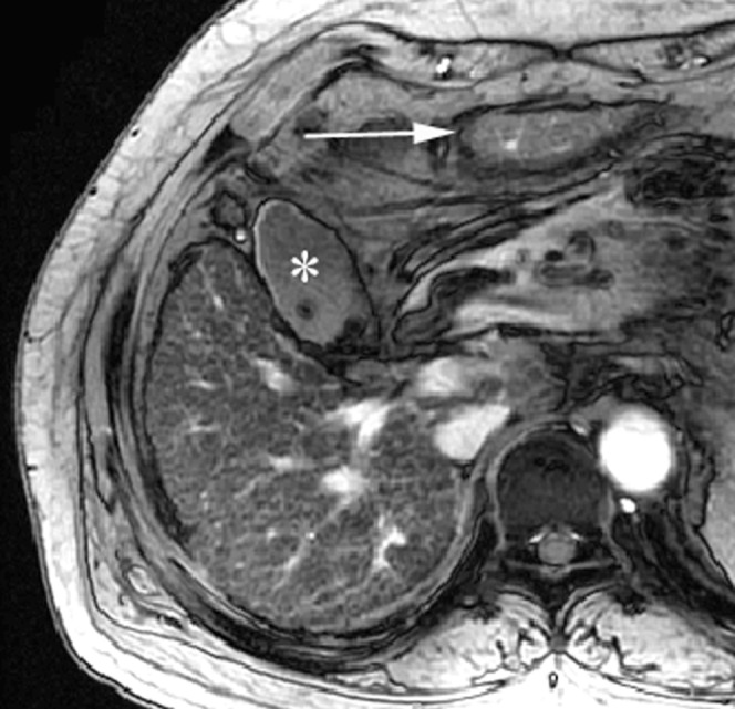
A 55-year-old woman with chronic viral hepatitis B categorised into Group 2 who was positive for the expanded gallbladder sign. Axial gradient-echo image (repetition time/echo time (TR/TE) = 160/5.8 ms) shows fine reticulations and innumerable nodules in the entire liver parenchyma (reticulation score = 2, nodularity score = 3); the inferior portion of the lateral segment (arrow) is demonstrated on the same plane as the gallbladder (asterisk) owing to the atrophy of the medial segment. The right posterior hepatic notch was not defined, and there were no portal hypertensive stigmata (not shown).