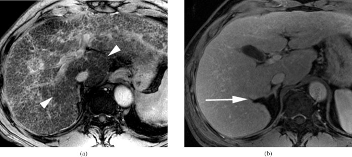
Figure 4.
A 59-year-old man with chronic viral hepatitis C categorised into Group 3 who was positive for the right posterior hepatic notch sign. (a) The entire liver parenchyma consists of fibrotic reticulations with variable thickness and micronodules, as seen on an axial gradient-echo image (repetition time/echo time (TR/TE) = 160/5.8 ms) (reticulation score = 4, nodularity score = 4). The caudate lobe is relatively spared from the fibrotic changes and shows darker signal intensity (arrowheads) than the remaining parenchyma. (b) The right posterior hepatic notch (arrow) is well defined on the axial contrast-enhanced three-dimensional gradient-echo image (TR/TE = 4.2/1.2 ms). The expanded gallbladder fossa sign was negative, and the spleen was enlarged up to 13 cm (not shown).

