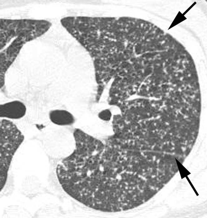
Figure 1.

Miliary tuberculosis in a 33-year-old woman not infected with HIV. A lung window of a transverse thin-section CT (1.0 mm section thickness) scan obtained at the level of the left upper lobar bronchus shows uniform-sized small nodules and micronodules randomly distributed throughout both lungs. Also note the subpleural and subfissural micronodules (arrows).
