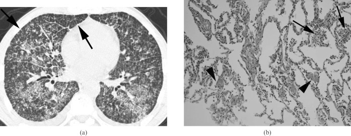
Figure 2.
Miliary tuberculosis presenting as acute respiratory distress syndrome in a 45-year-old man not infected with HIV. (a) A lung window of a transverse thin-section CT (1.0 mm section thickness) scan obtained at the level of the inferior pulmonary vein shows randomly distributed small nodules and micronodules with bilateral extensive ground-glass attenuation in both lungs. Also note the interlobular septal thickening (arrows) and intralobular interstitial thickening in both lungs. (b) A photomicrograph of a pathological specimen (haematoxylin and eosin staining; original magnification ×400) obtained with a transbronchial lung biopsy demonstrates poorly formed granulomas (arrows) in the alveolar wall and diffuse alveolar wall thickening and intra-alveolar fibrin deposition (arrowheads), suggesting an early stage of diffuse alveolar damage.

