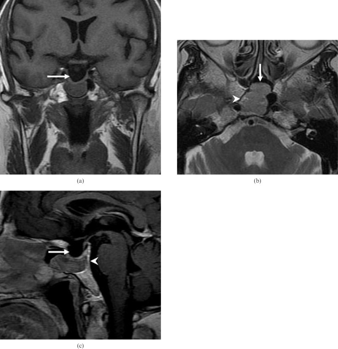
Figure 1.
Patient 2. (a) Coronal T1 weighted MR image shows a well-defined mass of isointense signal intensity in the sphenoid sinus with an intact sellar floor and empty sella (arrow). (b) Axial T2 weighted MRI shows a mass of intermediate signal intensity (arrow) with small foci of high signal intensity (arrowhead). Note the tumour compressing the adjacent clivus. (c) Sagittal contrast-enhanced MRI shows a mass of well-defined moderate heterogeneous enhancement. There is a thin gap between the superior margin of the tumour and sellar floor. Note the intacting sellar floor, mild remodelling of the adjacent clivus (arrowhead) and empty sella (arrow).

