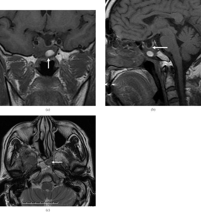
Figure 3.
(above) Patient 5 (had right nasal cavity endoscopic sinus surgery owing to a papilloma 10 years ago). (a) Coronal T1 weighted MR image shows heterogeneous sphenoid sinus ectopic pituitary adenoma with an area of high signal intensity (arrow), suggesting proteinaceous secretions. Note the presence of an empty sella and encasement of the right internal carotid artery. (b) Sagittal T1 weighted MR image shows an empty sella and intact sellar floor (arrow). Note the erosion of the adjacent clivus. (c) Axial T2 weighted MR image shows a sphenoid sinus mass of slightly high signal intensity with area of low signal intensity (arrow). Note the erosion of the adjacent clivus.

