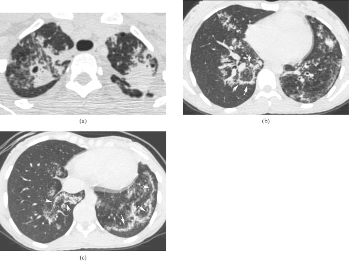
Figure 1.
A high-resolution CT scan showing patchy consolidations and ground-glass opacities in both lungs, with a cavitation in the right upper lobe (a). Opacities with central areas of ground-glass attenuation surrounded by denser crescentic consolidations of at least 2 mm thickness (reversed halo sign; arrows) are seen in the lower lobes (b,c). These areas are admixed with poorly defined nodules. No lymphadenopathy is evident.

