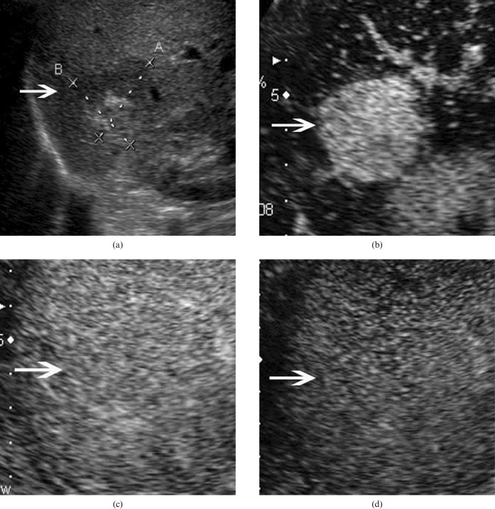
Figure 1.
Hepatic angiomyolipoma in a 42-year-old woman. (a) Baseline ultrasound scan shows a mixed echogenic lesion (arrow) 3.4 cm in diameter in segment 7 of the liver. Hyper- and hypoechoic portions are found in the lesion. (b) In the arterial phase of contrast-enhanced ultrasound, the lesion (arrow) shows homogeneous hyperenhancement 13 s after contrast agent injection. The lesion (arrow) shows isoenhancement in the (c) portal phase (100 s after contrast agent injection) and (d) late phase (130 s after contrast agent injection).

