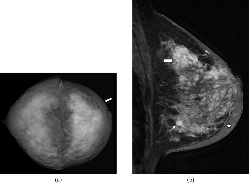
Figure 4.
40-year-old lactating woman, 9 months post partum, who presented with an enlarged, firm breast, inverted nipple and peau d'orange. Percutaneous ultrasound-guided biopsy revealed invasive ductal carcinoma and ductal carcinoma in situ (DCIS). Pre-operative imaging revealed bone and liver metastases. (a) Bilateral craniocaudal mammogram reveals extremely dense breast tissue. There is diffuse skin thickening (arrow) in the left breast. (b) Sagittal T1 weighted contrast-enhanced MRI demonstrates multiple masses and diffuse breast enhancement along with pronounced skin thickening (star) – findings are consistent with inflammatory breast carcinoma.

