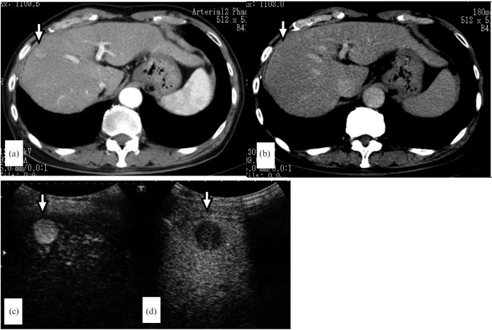
Figure 4.
A 69-year-old man with a 1.6 cm hepatocellular carcinoma in liver segment V. (a) The arterial phase of dynamic CT reveals a hypervascular tumour (arrow). (b) The portal phase of dynamic CT shows washout (arrow). (c) Contrast-enhanced ultrasound at the early vascular phase clearly shows hyperenhancement (arrow). (d) Contrast-enhanced ultrasound at the post-vascular phase shows hypo-enhancement (arrow).

