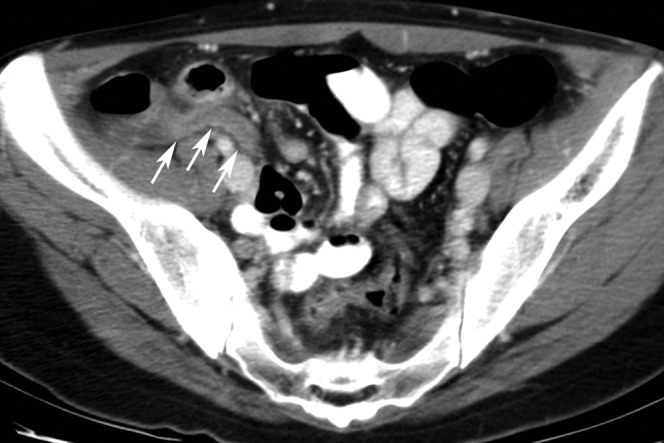
Figure 1.

A 27-year-old male patient with positive oral contrast and proven appendicitis correctly diagnosed by all readers. This axial CT scan shows a dilated appendix with wall thickening and increased enhancement of the wall (arrows) associated with mild peri-appendiceal stranding indicative of appendicitis. The lumen of the appendix does not contain positive oral contrast probably owing to inflammation.
