
Figure 5.
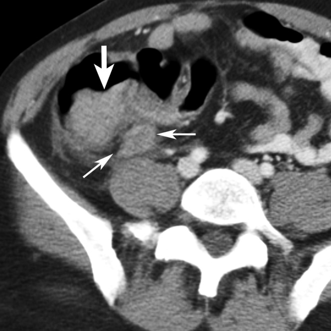
A 45-year-old male patient with positive oral contrast and a false-positive diagnosis of appendicitis by three of the five readers (Reader 3, a fellow; Readers 4 and 5, two first-year residents). The appendix appears enlarged (thin arrows) and the cecal wall is nodular and thickened (thick arrows). At surgery a cecal carcinoma with microperforation was found, which might have caused the apparent appendiceal inflammation. The positive oral contrast did not reach the cecum by the time of the scan.