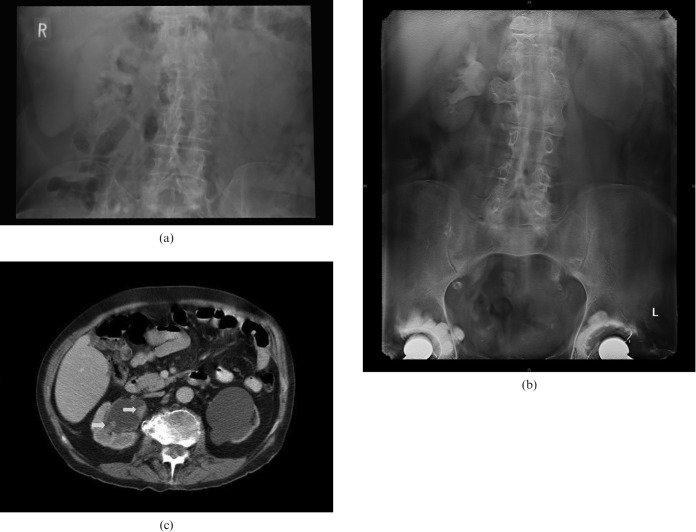
Figure 2.
(a) Traditional intravenous urography giving little information about the left kidney and non-diagnostic for the right. (b) Digital tomosynthesis obtained contemporaneously on the same patient reveals two foci of transitional cell carcinoma in the pelvis of the right kidney, and hydronephrosis with a thin rim of renal cortex on the left owing to longstanding pelvi–ureteric junction obstruction. Note the exquisite detail seen in the transverse vertebral process at the same level. (c) The same features shown on CT in this patient, for correlation. Arrows show transitional cell carcinoma foci.

