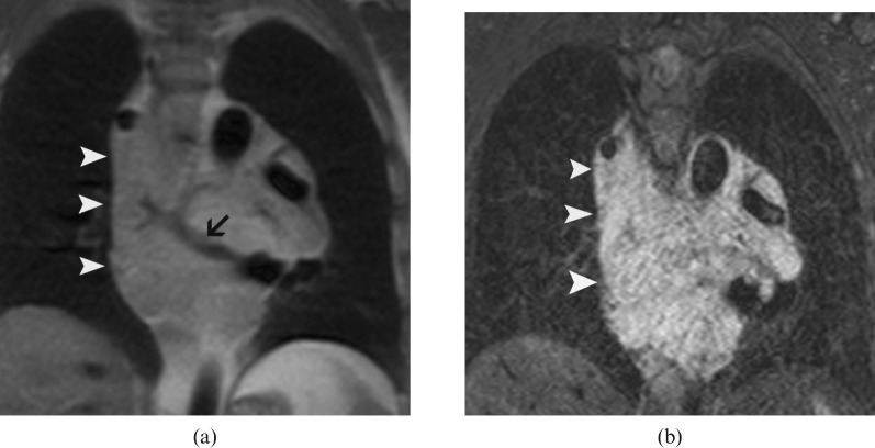
Figure 1.
Coronal MR images of the mediastinum show the heterogeneous, infiltrative malformation in the middle mediastinum. The mass (arrowheads) is isointense on T1 weighted black blood images (a) and hyperintense on short tau inversion recovery images (b). A vascular structure is seen as flow void within the mass (arrow) (a).

