
Figure 1.
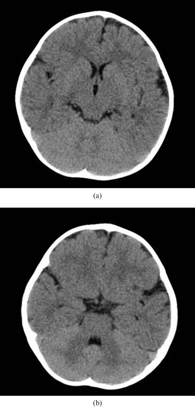
Multidetector CT images representing the levels of the brain used for diagnostic image quality assessment: (a) the upper level of the brain, showing the lateral ventricles and the basal ganglia, and (b) the lower level of the brain, including the posterior fossa at the level of the fourth ventricle. The patient is female, 15 months old, and was scanned with the parameters according to Table 1.